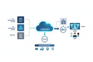
Forget bulky film jackets and lost images! Modern radiology thrives on PACS, a digital powerhouse that stores and organizes scans from CTs, MRIs, X-rays, and even ultrasounds.
Think of it as a Netflix for medical images, allowing doctors to access and analyze a patient's entire visual history instantly. This streamlined approach speeds up interpretation, empowers collaboration, and contributes to more accurate diagnoses and better patient outcomes.
Let's dive deeper and explore how PACS goes beyond storage to revolutionize radiology!
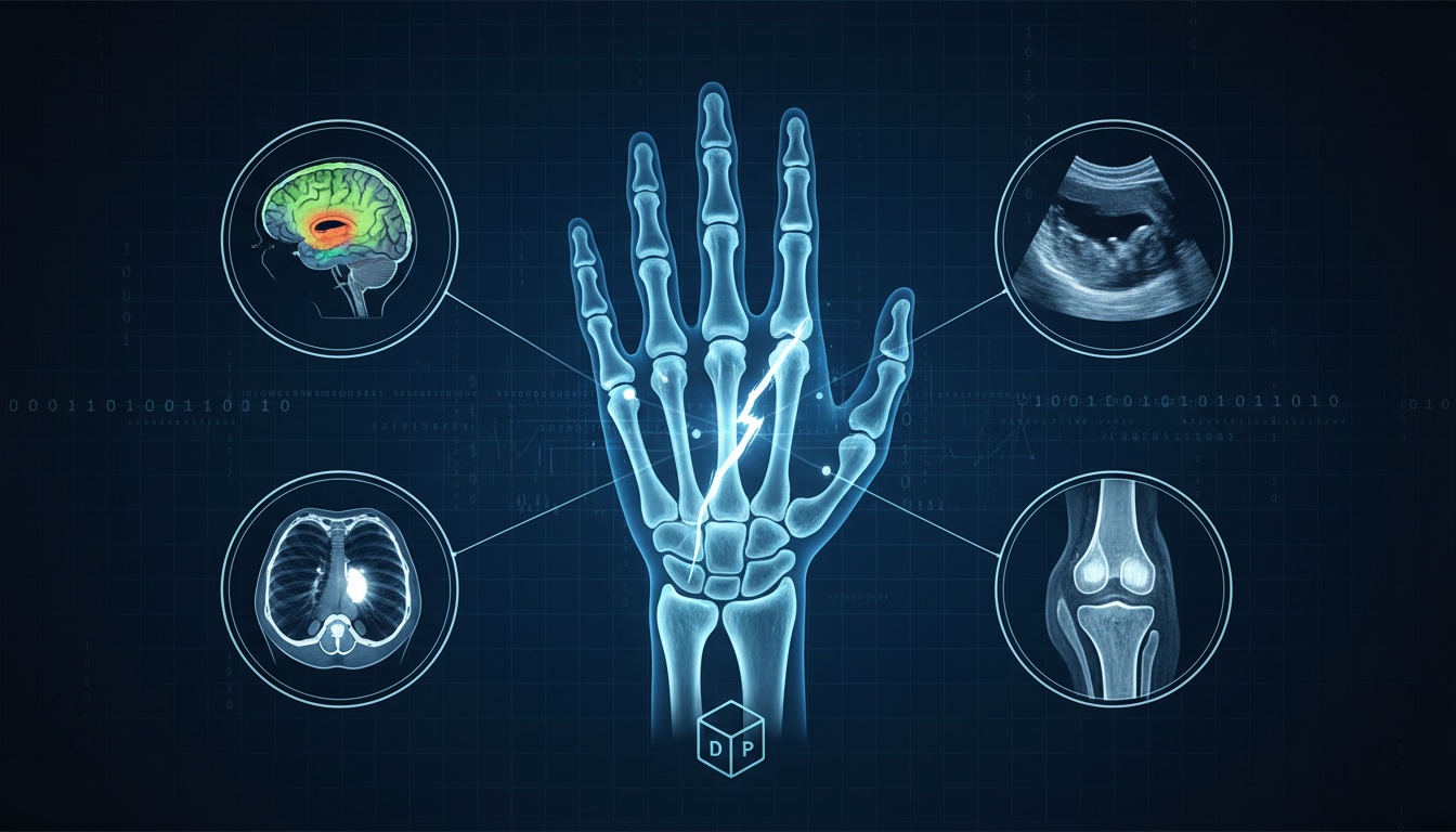
A broken bone is more than a painful inconvenience; it can signal significant injury and potential complications.
That's why radiologists rely on PACS to visualize fractures with unparalleled accuracy, empowering them to provide the precise diagnoses needed to guide treatment decisions.
The humble X-ray remains a cornerstone of fracture detection. With PACS, X-ray images transform from static films to dynamic digital tools. Radiologists can zoom into areas of interest, revealing subtle fracture lines that might be missed on traditional film.
Adjusting brightness and contrast can be crucial for visualizing fractures in dense areas or patients with varying body compositions.
When a simple break turns complex, CT scans become invaluable. PACS platforms truly shine with CT, handling massive datasets and allowing radiologists to slice through the anatomy layer by layer. This reveals the precise location of a fracture, its extent, displacement, and any associated bone fragmentation.
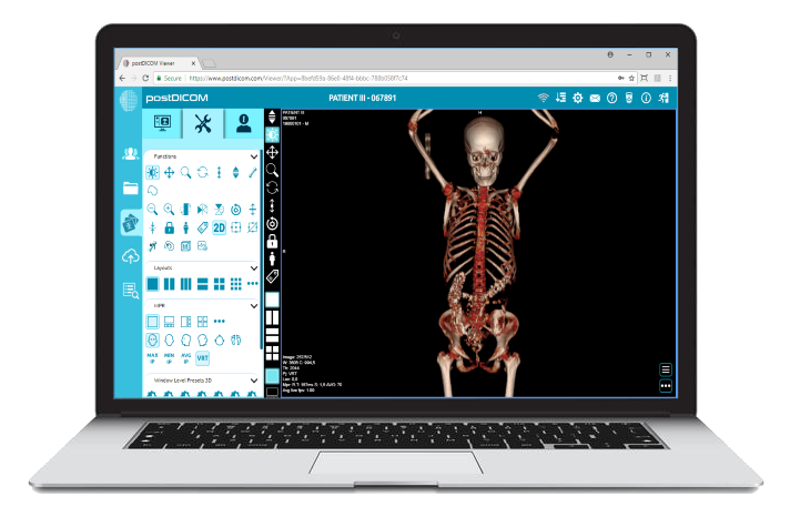
Advanced PACS solutions often offer 3D reconstruction capabilities. With a few clicks, your doctors can rotate a 3D model of a fractured bone, gaining a spatial understanding that's impossible with flat images alone. This is particularly helpful for surgeons planning complex fracture repairs.
While bone shines brightly on X-rays and CT, MRI visualizes soft tissues. This can be critical in assessing the extent of ligament, tendon, or muscle damage associated with a fracture.
PACS allows seamless viewing of these MRI scans alongside other modalities, providing a comprehensive picture of the injury.
Beyond image display, PACS systems offer tools that directly enhance fracture analysis:
Measurements: Precise measurement tools allow for accurately assessing fracture displacement and angulation, which is crucial for determining treatment methods.
Comparison: Easily accessing prior scans on a PACS lets radiologists track fracture healing, spot complications, or assess hardware placement post-surgery.
Collaboration: PACS empowers specialists to consult quickly on complex cases. An orthopedic surgeon can discuss a 3D fracture reconstruction with the radiologist in real-time, no matter their location.
PACS ensures that every pixel of a patient's imaging data is utilized to its full potential, from the emergency room to the orthopedic surgery suite.
The ability to precisely manipulate, analyze, and share images is critical for diagnosing fractures and understanding the full scope of an injury.
Internal bleeding can be a ticking time bomb, every second without a diagnosis, increasing the risk of serious complications or even death. Radiologists rely on CT scans and PACS to detect even subtle signs of bleeding, potentially saving lives.
CT scans are exceptionally powerful for visualizing blood within the body. The bleeding appears brighter than surrounding tissues, making it easier for a trained radiologist to spot.
The speed of CT image acquisition is also a significant advantage – a patient with suspected internal injuries can be scanned in minutes, allowing for rapid diagnosis and intervention.
PACS takes CT bleeding detection to the next level. Here's how:
Subtle Signs: Radiologists can zoom into areas of concern on a CT scan, potentially revealing small areas of bleeding that might be missed on a static image. Manipulating image brightness and contrast can also highlight subtle changes in density that signal blood presence.
The Bigger Picture: PACS allows radiologists to review multiple CT scans side-by-side. This is crucial for pinpointing the source of bleeding, such as a ruptured organ or damaged blood vessel.
Quantifying Blood Loss: Some PACS solutions offer tools to estimate the volume of blood present, providing doctors with valuable information for determining the severity of the injury.
Internal bleeding can evolve, either worsening or resolving. With PACS, radiologists have an invaluable patient imaging history timeline. By comparing past CT scans to the most recent ones, they can track the progression of bleeding. This is essential for:
Monitoring Stability: Conservative management might be appropriate if bleeding appears stable on serial scans.
Catching Complications: If bleeding worsens, rapid intervention may be needed.
Surgical Guidance: PACS helps surgeons visualize the precise location and extent of bleeding, aiding in planning lifesaving procedures.
While image visualization is at the heart of PACS, the additional benefits it offers in the context of internal bleeding are profound:
Speed: PACS ensures CT scans are available to ER physicians and surgeons in near real-time, accelerating decision-making in critical situations.
Remote Access: In trauma cases, cloud-based PACS lets specialists consult from afar, expanding the expertise available to the patient.
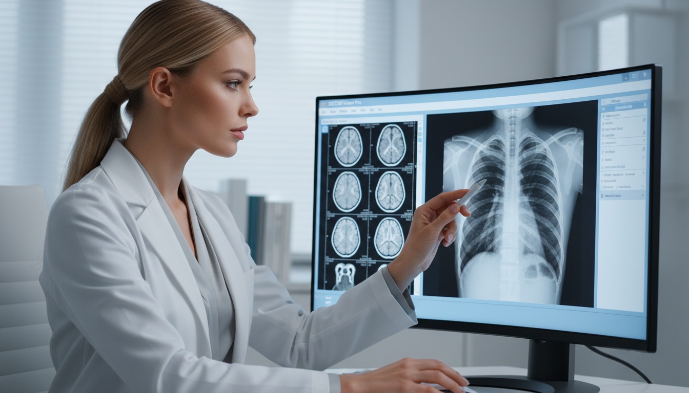
The lungs are complex organs susceptible to a vast array of diseases. Detecting these conditions, from tiny nodules to widespread pneumonia, often relies heavily on imaging paired with the power of PACS.
Lung nodules, small growths within the lung, can be an early warning sign of cancer or other serious diseases. CT scans are excellent for detecting them, but some nodules are incredibly subtle.
PACS provides the tools radiologists need to find these potentially dangerous abnormalities.
Zooming into specific areas of the lung reveals details impossible to see in a traditional image. PACS's ability to fine-tune the brightness and contrast lets radiologists highlight faint nodules that might otherwise blend into the background, potentially making the difference between early and late-stage diagnosis.
Pneumonia, an infection of the lungs, appears on chest X-rays and CT scans. PACS makes interpreting these images much easier. Adjusting image settings can reveal subtle areas of inflammation masked on static films.
Comparing previous chest imaging on PACS lets radiologists determine if a suspected pneumonia is new or represents a chronic condition.
PACS aids in visualizing a wide range of lung pathologies, including:
Pulmonary Embolism (Blood Clots): PACS optimized for CT image viewing helps detect clots in lung arteries, a potentially life-threatening condition.
Emphysema: Analyzing CT scans on PACS assists in quantifying lung damage from emphysema, aiding disease staging and treatment decisions.
Interstitial Lung Disease: The subtle patterns seen with these diseases are better visualized with PACS's fine adjustment capabilities.
The advantages of PACS for lung imaging go beyond simple viewing:
Measurements: PACS often includes tools to measure nodules, track their growth over time (crucial in cancer monitoring), and assess the extent of pneumonia.
3D Reconstructions: Some PACS platforms offer 3D modeling of the lungs using CT data, adding a new dimension to understanding complex diseases.
AI Integration: Emerging PACS solutions integrate artificial intelligence algorithms, which can highlight suspicious nodules or automatically quantify areas of pneumonia.
 - Created by PostDICOM.jpg)
When it comes to diagnosing conditions of the brain and spinal cord, MRI is king. It offers unparalleled detail of soft tissues and reveals abnormalities that would be invisible on CT scans.
PACS takes MRI visualization to the next level, unlocking insights essential for accurate diagnosis and treatment planning.
Brain tumors come in a bewildering variety of types and locations. PACS helps radiologists visualize them with precision on MRI scans.
Fine adjustments of brightness and contrast can highlight a tumor's boundaries, helping differentiate it from normal brain tissue.
Advanced PACS platforms often offer specialized image processing tools for further analysis of brain tumors, extracting information about their blood supply or aggressiveness.
Multiple sclerosis (MS) is characterized by lesions scattered throughout the brain and spinal cord.
PACS allows for meticulous comparison of MRI scans over time. This is crucial, as the appearance of new lesions or changes in existing ones can guide treatment decisions for patients with MS.
The spinal cord is a delicate and complex structure. Injuries, inflammation, or degenerative conditions can cause subtle changes that are easily missed without sophisticated image analysis.
PACS empowers radiologists to zoom into the smallest details of the spinal cord on MRI. Specialized tools may allow for precise measurements or 3D reconstructions, aiding in visualizing even the most elusive abnormalities.
Beyond basic visualization, here's what sets PACS apart for neurological imaging:
Multi-Sequence Viewing: MRI generates different image types (T1, T2, FLAIR, etc.). PACS lets radiologists examine all these sequences seamlessly, each offering unique insights into specific tissues.
Brain Mapping Tools: Some PACS platforms specialize in neurological imaging analysis, overlaying functional MRI data onto anatomical scans to pinpoint areas of brain activity. This is revolutionizing neurosurgery and research.
Advanced Quantification: Certain solutions go beyond simple viewing, using algorithms to automatically segment tumors, measure brain structures, or even detect subtle MS lesions a human eye might miss.
Neurological diagnosis is high stakes. Missed or delayed findings can have life-altering consequences for patients. PACS ensures that not a single pixel of precious MRI data goes unexamined.
This empowers neurologists and radiologists with the comprehensive view they need to detect tumors at their earliest stages, accurately assess MS progression, and pinpoint spinal cord conditions that require urgent intervention.
While not every PACS is equal, solutions like PostDICOM that emphasize advanced image processing and collaborative features are particularly beneficial for facilities handling complex neurological cases.
Ultrasound has seen a remarkable rise in recent years. Its safety, accessibility, and real-time imaging capabilities are transforming many areas of medicine.
But with this expanded use comes a new challenge: storing and managing the vast amounts of ultrasound data generated. That's where PACS steps in once again.
Let's take a quick tour of how ultrasound reveals what other imaging modalities can't:
Gallbladder Stones: Ultrasound is the gold standard for detecting these, often catching stones too small to see on other scans.
Fetal Imaging: The most well-known use, ultrasound, allows for incredible views of a developing baby, assessing growth and detecting abnormalities.
Blood Clots: Ultrasound can visualize clots in the legs (DVTs), a potentially life-threatening condition.
Thyroid Nodules: Small thyroid nodules are easily visualized with ultrasound, aiding in biopsy and cancer diagnosis.
Guided Procedures: Ultrasound offers real-time guidance for needle biopsies, fluid drainages, and more, increasing accuracy.
Like CT and MRI, ultrasound exams generate a huge number of images and video clips. PACS has become essential for storing and organizing this data.
Modern PACS solutions are designed to handle the unique file types and workflow associated with ultrasound.
This translates to some significant advantages:
Side-by-Side Comparisons: Review a new fetal ultrasound alongside prior ones, precisely tracking growth. Or, compare a gallbladder scan to an older one, assessing for changes over time.
Easy Access for Specialists: PACS makes ultrasound images easily accessible to obstetricians, cardiologists, surgeons, and specialists who need information for diagnosis and treatment planning.
Remote Review: Cloud-based PACS lets physicians review ultrasound scans off-site, which is excellent for urgent consults or getting second opinions quickly.
Teaching & Learning: PACS facilitates the creation of ultrasound image libraries to educate medical students or residents.
Initially, DVDs or basic local servers often handled ultrasound image storage. PACS elevates the process, centralizing storage and unlocking powerful viewing capabilities.
By integrating ultrasound seamlessly with other imaging modalities, PACS creates a comprehensive picture of a patient's health.
When considering a PACS solution, it is essential to choose one optimized for ultrasound's unique file formats and fast enough to stream video clips smoothly.
This ensures your medical teams can get the most out of the incredible images ultrasound provides, leading to better diagnosis, treatment, and care across numerous specialties.
PACS gives radiologists the tools to see what was once invisible, from X-rays to ultrasounds. It breaks down barriers, allowing specialists to collaborate effortlessly on even the most complex cases.
With reliable PACS, images are always a click away, ensuring that fast, accurate diagnosis becomes the norm. Choosing the right PACS provider, like PostDICOM, is the key to unlocking the full potential of this transformative technology.
|
Cloud PACS and Online DICOM ViewerUpload DICOM images and clinical documents to PostDICOM servers. Store, view, collaborate, and share your medical imaging files. |