A CT of the brain is a noninvasive diagnostic imaging procedure that uses special X-rays to produce horizontal or axial images (often called slices) of the brain. The CT scan of the brain provides more detailed information about brain tissue and the brain’s overall structure than standard X-rays. The detailed structure thus provides more data related to injuries or diseases of the brain.
Besides Brain CT and X-rays, there are other procedures that are used to diagnose brain injuries or disorders. The procedures include Magnetic Resonance Imaging (MRI), Positron Emission Tomography (PET), and Cerebral Arteriogram.
During the brain scan, the X-ray beam moves in a circle around the body, taking different brain views from different angles. The information recorded by X-ray is then sent to a computer that interprets the data and displays them in a 2D form on the monitor.
The CT scans can be done with or without contrast. Contrast means a substance that is taken via the mouth or injected with an intravenous line that causes a particular tissue or organ to be able to see more clearly. Contrast examinations are used for studying the parts of your body more closely. The examination requires fasting for a certain period before the procedure takes place.
 - Created by PostDICOM.jpg)
A brain CT scan helps assess the brain for diagnosing tumors, injuries, intracranial bleeding, brain hemorrhage, structural deformities, etc. The CT scan is conducted when other means of examination aren’t conducive.
The CT scan of the brain can be used to evaluate the effects of treatment on brain tumors and to detect any clot formation post strokes. The scans also provide guidance for brain surgery or undergoing tissue biopsies.
1. Your diagnoser may ask you to change into a patient’s gown. You need to wear it and remove all jewelry from you.
2. Next, you will be asked to sign a consent form explaining all the side effects associated with contrast media injected through an intravenous or IV line.
3. For brain CT with contrast, you need to fast for at least 3 hours prior to the CT. However, if you do not need a contrast, you can eat, drink and take your prescribed medicines before your exam.
4. If you have diabetes, you should eat a light breakfast or lunch 3 hours before the scheduled scan. Depending on your diabetic medication, you may be asked to discontinue using them 48 hours after the CT.
5. Whether you’re a diabetic patient or not, you can take your prescribed medications as usual unless your doctor tells you not to.
Step 1: If you are given contrast, an IV line will be started in the arm or hand to inject the contrast media. You could be given oral contrast too. The oral contrast comes in a liquid form which is given for patients to swallow.
Step 2: You will be lying on a scan table that slides into a large machine with a circular opening. The pillows and straps can be used to prevent movement during the process.
Step 3: The CT operator will be in another room with the scanner controls. However, they will keep an eye on you through the window. You will be able to hear your diagnoser with the help of speakers that are installed inside the scanner. This will allow two-way communication between you and them.
Step 4: When the diagnoser operates the scanner, it will start rotating around you, and X-rays will pass through the body for short amounts of time. You can hear clicking sounds, but don’t panic. This is normal.
Step 5: As the scanner begins to rotate around you, X-rays will pass through the body for short amounts of time. You will hear clicking sounds, which are normal.
Step 6: As the scanner rotates around you, the X-rays will pass through the body for a minimal amount of time. The rays absorbed by your body will be detected by the scanner and transmitted to the computer.
Step 7: If contrast is used for your procedure, you may feel some effects when the media is injected into you. You may feel a flushing sensation or a salty or metallic taste in your mouth. Some even experience mild headaches or nausea. But don’t worry; these effects are short-lived. If you encounter discomfort or breathing difficulties, notify your diagnoser.
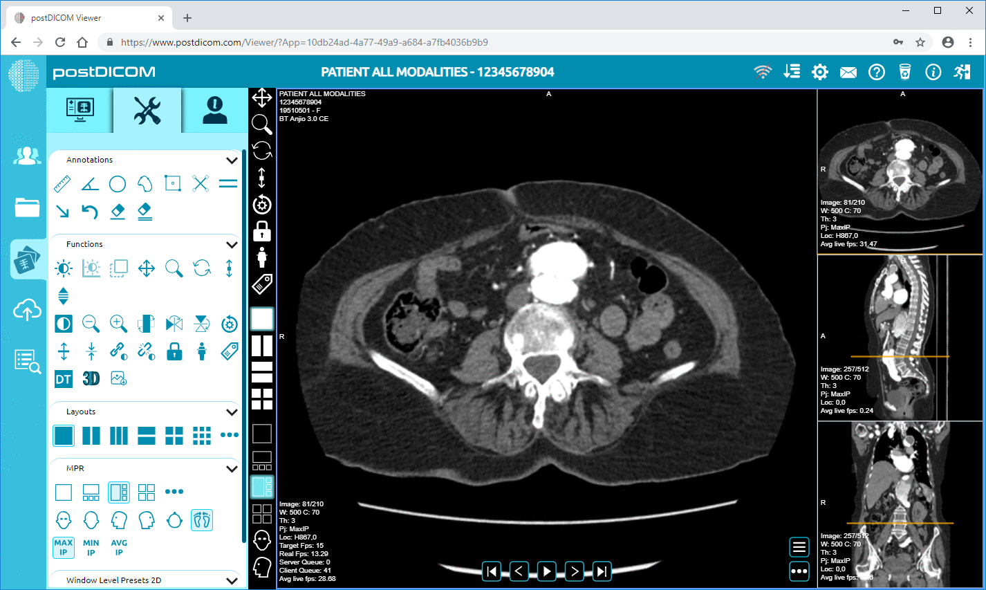
Step 8: After the procedure is completed, you will get up from the scanner. If they used an IV on you, then the line will be removed. The computer will transform the information into a DICOM image. The radiologist can either give you documents physically or can store them in PACS and give you access to the reports by sharing a link.
Hypodensity is an abnormality found on CT scans. It means possible open spots or fluid-filled spots where they appear darker than the other parts of an organ or a tissue.
On a head hypodensity CT scan, the areas represented in darker hues are hypodense. The appearance of the dark area can signify abnormalities like strokes, edema, masses, air or fat abnormalities, etc. The radiologist will be able to provide a diagnosis or some possibilities.
On a chest CT scan, the darkest areas on the lung are hypodense. These areas can contain cysts or air trapping. Masses, cysts, and lymph nodes can be considered hypodense in the mediastinum. If blood vessels have hypodense, it means there’s a clot or occlusion present. Hypodense on the chest wall or ribs may be bone lesions.
The CT of the pelvis and abdomen will represent similar outcomes. The hypodense areas may appear darker because of cysts or masses. Hypodense in the bowel and colon may be air, masses, stool, fat, or other deformities.
Hypodense on the bones may indicate a bone lesion, such as a Lytic lesion. A lytic lesion is darker than the bone and can be described as hypodensity. Hypodensity in the bones may also represent normal marrow or structure of the bone.
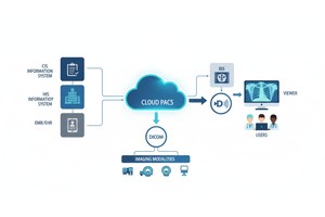
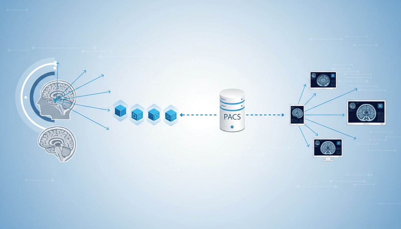
The patient’s data extracted by the CT scanner are sent to the PACS through the Router. All data acquired by the scanner are then stored in the PACS.
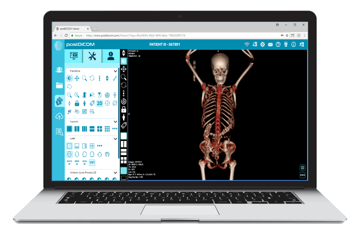
In PostDICOM’s case, you can use our cloud-based PACS to store, view, and retrieve the data with great ease. A cloud-based PACS system expands the computer network, allowing a more comprehensive range of devices to act as display stations. Any HTML5-compatible device can access a cloud-based DICOM viewer online.
If you haven’t already, you should switch to PostDICOM’s cloud PACS system. Here are the perks of switching:
It’s cheaper than regular PACS
It offers managed storage
It takes up less space
It is low maintenance
It’s safer to use
It provides easy access to images
It makes collaboration convenient
PostDICOM offers cloud-based PACS and DICOM viewers with a free trial period. After the trial period ends, you can choose to expand the cloud storage and get extra features just by paying a meager subscription fee, starting from 79.99.
|
Cloud PACS and Online DICOM ViewerUpload DICOM images and clinical documents to PostDICOM servers. Store, view, collaborate, and share your medical imaging files. |