Blood clots may be significant if they block the usual flow of blood and, if they move to vulnerable areas of the body. That is why medical imaging might be used to help doctors quickly find a clot and to decide on the best treatment. A common query that many patients have is, can MRI detect blood clots?
Yes. The short answer is, it can. Because an MRI can find blood clots, especially in the deep veins, brain, abdomen, pelvis and other soft tissue areas with blood vessels, it is very helpful. An MRI is not always the first choice for imaging. For emergencies, a CT scan or ultrasound might be faster and more convenient. The type of imaging used will depend on where the clot is found, what imaging is available, and the patient's medical history.
Once you understand the potential advantages of MRI imaging in the diagnosis of vascular problems, it's easier to appreciate why some may be recommended over others.
Yes. MRI can diagnose many blood clots and circulatory problems. It can be very useful to evaluate brain clots, clots in the pelvis, clots in the abdomen, clots in deep veins and some chronic vascular disorders. However, depending on the symptoms, suspected location and urgency, ultrasound or CT scans are often the first choices.
Magnetic Resonance Imaging (MRI): A non-invasive imaging technique which uses powerful magnets and radio waves to produce a very detailed image of the structures within the body. MRI does not employ ionizing radiation, as does computed tomography (CT) or simple X-rays.
MRI is particularly useful when doctors require clear images of soft tissues, blood vessels, organs, nerves and the brain. Contrast dye can be injected to make it easier to see blood flow and the blood vessels' shapes.
MRI can be very helpful in the evaluation of some blood clots due to the excellent tissue contrast achieved with this technique.
MRI can show either direct or indirect evidence of clots forming. Radiologists may view:
• Blocked Or Narrowed Blood Vessels
• A Lack Of Or Abnormal Blood Flow
• Inflammation And Swelling Around The Involved Tissues
• Damage To Tissues Due To Poor Circulation
• Clot Material Directly In The Vessel
Arteries and veins are frequently examined in greater detail using special MRI techniques like MR Angiography (MRA) and MR Venography (MRV). These are used to determine if the circulation is normal or blocked.
 - Created by PostDICOM.jpg)
MRI is not used for every clot, but in several cases is a valuable test.
MRI is frequently ordered if there is a suspicion of stroke. When a blood clot prevents blood from flowing to part of the brain, MRI is able to sometimes identify the injury at an early stage, and assist the doctor in determining the amount of damage.
MRI may also be useful in differentiating various causes of neurological symptoms.
Sometimes a blood clot develops in the veins that drain the blood from the brain. Cerebral venous sinus thrombosis (CVST) is the name for this condition. When this condition is suspected MR Venography is often employed.
There are some deep veins in the pelvis or abdomen which are more challenging to evaluate by ultrasound. MRI may be better suited to visualize these areas, especially if symptoms don't go away and previous imaging is not definitive.
MRI also may be helpful if symptoms persist over time, if previous tests leave questions unanswered, or if the doctor requires more information before making treatment decisions.
While MRI is very effective, it is not always the first test used to image blood clots.
In the case of suspected deep vein thrombosis (DVT) of the legs, ultrasound is often the first treatment option. Faster, more readily available, painless, and non-radiating.
In emergency situations, CT scans (particularly CT angiography - CTA) are often used. They are typically used when the patient has suspected pulmonary embolus (blood clot in the lungs) or urgent blockage of the vessels is suspected as they can provide the result quickly.
In situations where ultrasound can't be used, or when doctors require more detailed soft-tissue pictures, detailed vascular pictures, or MRI is the preferred method.
| Imaging Test | Common Use for Clots | Key Advantage |
| Ultrasound | Leg DVT | Fast, accessible, no radiation |
| CT / CTA | Lung clot, emergency vascular problems | Rapid imaging |
| MRI / MRA / MRV | Brain, pelvis, abdomen, complex cases | Excellent soft-tissue detail |
There are advantages to each imaging tool. There is no one scan which is better than another; it depends on the clinical situation.
Although MRI is very valuable, no imaging examination is completely accurate. The accuracy will depend upon the site of the clot, the time of the scan, the quality of the scan and the technique employed. Further imaging or repeat testing may be ordered if symptoms continue to be an issue.
Yes, MRI will pick up DVT in many instances. But ultrasound is the most frequently performed test in the legs when a clot is suspected; it is also quicker, cheaper and very effective.
MRI may be indicated when:
• Ultrasound Results Are Unclear
• The Clot May Be Higher In The Pelvis
• More Anatomical Detail Is Needed
• A Complex Or Recurrent Case Is Being Investigated
Typically not as a first test. When there is a suspicion of DVT in the leg, ultrasound is more readily available, quicker, and very effective. MRI is more frequently used when ultrasound is inconclusive or deeper veins are to be seen.
Yes. MRI is one of the most useful methods used to identify these types of issues caused by clots in the brain. It can be used to assess:
• Blocked Blood Flow Stroke (ischaemic Stroke)
• Cerebral Venous Sinus Thrombosis (c.v.s.t.)
• Damage To Tissue Due To Low Blood Flow
• Secondary Swelling Or Complications
As for the stroke treatment, the speed is of the essence, so in the emergency department, the CT scan is carried out first, followed, if applicable, by MRI.
In some instances MRI has certain advantages:
• No Ionizing Radiation
• Excellent Soft-tissue Contrast
• Strong Detail For Brain And Vascular Imaging
• Helpful In Deeper Anatomical Regions
• Useful When Previous Imaging Is Inconclusive
The advantages of MRI make the modality an important tool in the current vascular imaging toolbox.
MRI also has some practical restrictions:
• Longer Scan Times Than Ct.
• Not As Readily Accessible In Certain Emergency Situations
• Increased Cost For Many Health Systems
• Not Suitable For All Unstable Patients
• Some Patients Cannot Undergo Mri Because Of Certain Implants Or Severe Claustrophobia
That is why doctors make their decisions on imaging based on the individual case.
Any signs of a blood clot should not be overlooked. If you have symptoms, you may need immediate medical evaluation, such as:
• Sudden Leg Swelling
• Calf Pain/tenderness
• Chest Pain
• Shortness Of Breath
• Sudden Weakness Or Numbness.
• Trouble Speaking
• Severe Unexplained Headache
• Sudden Loss Of Vision Or Balance
Doctors use imaging for specific symptoms, need for urgency, and overall risk factors.
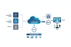
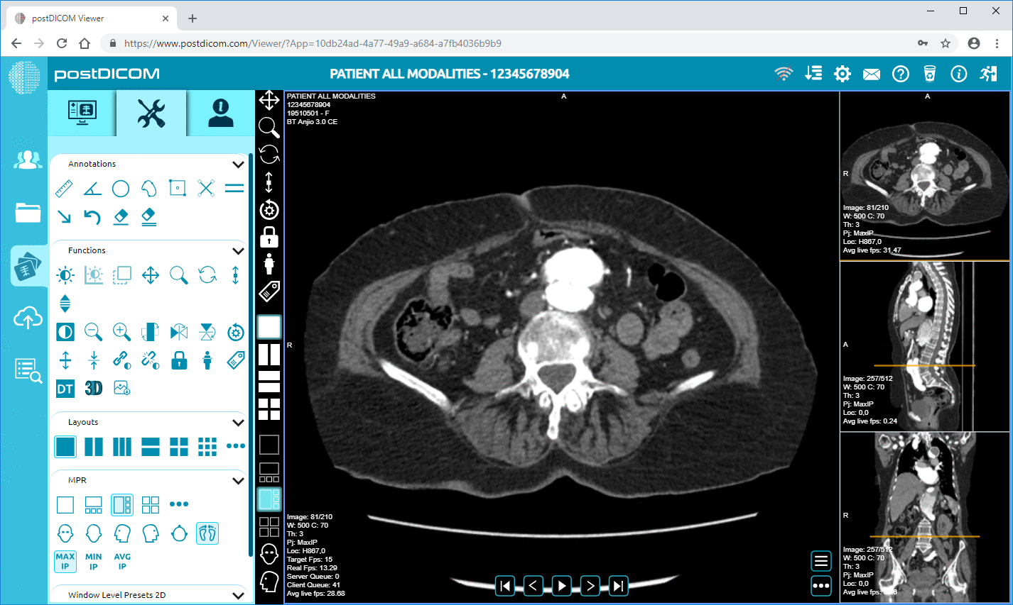
Once imaging has been done, gaining access to scans can facilitate team collaboration. When efficient workflows are required, medical images can be viewed, shared, and reviewed remotely, as do many modern cloud-based DICOM platforms like PostDICOM.
This can be particularly helpful if the specialists require timely access to MRI studies at multiple sites.
Treatment will be determined by the size, location and severity of the blood clot, if found by imaging. Depending on the case, blood thinners, monitoring, emergency procedures, and specialist follow up may be recommended.
Prompt diagnosis is essential as an untreated clot can be harmful or fatal if it prevents blood flow or moves to the lungs or other vital organs.
This really depends on where the blood clot's how bad the situation is. The doctor will usually use a CT scan in an emergency because it is fast and easy to get. Doctors might use an MRI if they want to look closely at the soft parts of the body or do advanced tests on the blood vessels.
If someone thinks a person may have a blood clot in the lungs, the doctor will usually do a computed tomography angiogram of the lungs first. MRI may be used, but it is not the first test in emergency cases.
No, MRI does not use radiation that can hurt you. Instead, it uses magnets and radio waves to make detailed pictures of your organs, blood vessels, and soft tissues.
No test is perfect. How well the test works depends on where the blood clots when the scan is done, how good the pictures are, and what method is used. If you are still having symptoms, the doctor may want to do tests or repeat the scan.
The time it takes will vary depending on which part of the body is being scanned and what kind of scan it is. Depending on the type of MRI, it could take 20 to 30 minutes. Even longer for the more complicated tests.
So, can MRI detect blood clots? Yes, MRI is very good at detecting blood clots and blood flow problems. In the brain, pelvis, abdomen, and blood vessels, especially, this is one tool doctors use.
All three tests, ultrasound, CT, and MRI, are important. Depending on where the blood clot's how bad the situation is, and what the doctor needs to know, the best test will be different.
Doctors can quickly and easily review test results with systems and secure platforms like PostDICOM, helping them make faster, more informed decisions about MRI and other tests, such as CT and ultrasound.
|
Cloud PACS and Online DICOM ViewerUpload DICOM images and clinical documents to PostDICOM servers. Store, view, collaborate, and share your medical imaging files. |