If doctors require a close-up view of the heart, they can use cardiac MRI or cardiac CT. Both are advanced imaging tests, but for different clinical indications.
The cardiac imaging test to use depends on whether the physician needs to assess the coronary arteries, the heart muscle, or cardiac function as a whole.
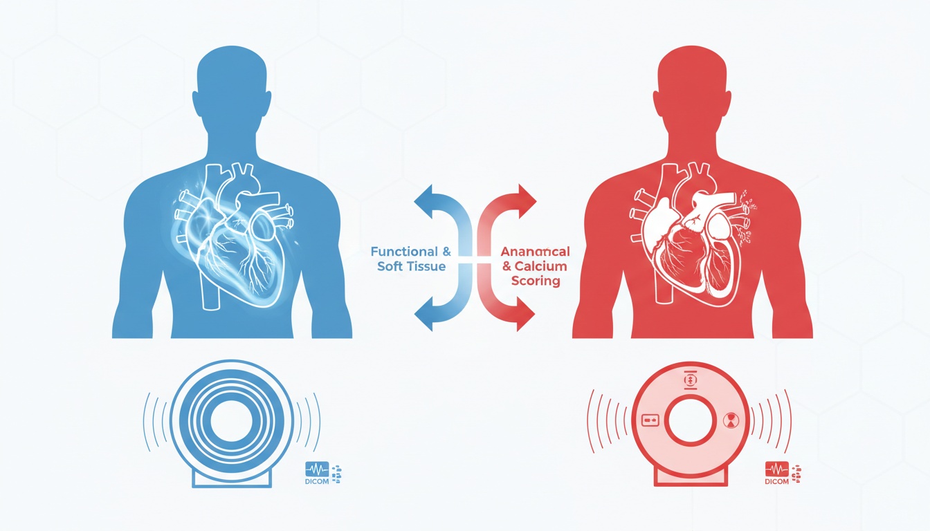
Cardiac CT typically offers a superior evaluation of the coronary arteries, the narrowing of blood vessels, and calcium deposits and plaques. Cardiac MRI is typically superior for things such as assessing function, inflammation, scarring, tissue damage, and viability.
Not all scans are created equal. The appropriate one is based on the patient's symptoms, medical history, and the question the physician wants answered.
This guide to CT and MRI imaging provides a general overview of the differences between imaging modalities to readers who are interested in knowing more about how these two imaging methods work.
Cardiac CT scans are used to produce detailed images of the cross-sections of the heart, using X-rays and computer processing. It is most commonly used in coronary CT angiography (CCTA), a procedure that helps doctors examine the coronary arteries that carry blood to the heart.
Cardiac CT may be done when physicians wish to evaluate for:
• Coronary Artery Disease
• Plaque Buildup
• Calcium Deposits
• Narrowed Or Blocked Coronary Arteries
• Chest Pain Related To Possible Heart Disease
• Heart And Blood Vessel Anatomy Before Procedures
A CT scan can help doctors rapidly and in detail examine the coronary arteries. Cardiac CT, however, involves ionizing radiation and may require iodinated contrast; therefore, renal function and a history of contrast allergy should be taken into account.
Cardiac MRI (magnetic resonance imaging) is a specialized MRI technique that is used to create detailed images of the heart. Unlike CT, MRI doesn't rely on the use of ionizing radiation.
Cardiac MRI may be useful for assessing damage to the heart muscle, chamber size, cardiac valves, blood flow, inflammation, and scar tissue after a heart attack. It can also be used to diagnose diseases such as myocarditis, cardiomyopathy, congenital cardiac disease, pericardial disease, and idiopathic heart failure.
Tissue characterization is the greatest advantage of cardiac MRI. This can reveal not only the heart's shape and action but also those of the myocardium (heart muscle). This is useful for a doctor to determine if the heart muscle is inflamed, scarred, weak, or enlarged.
The disadvantage of MRI is that it typically takes longer than CT. Other patients may also find it uncomfortable because they must remain still for a longer period in the scanner. Some newer cardiac devices are compatible with MRI, while for some patients who have older devices or implants, MRI may not be an option.
| Comparison Point | Cardiac CT | Cardiac MRI |
| Best for | Coronary arteries, plaque, calcium, vessel narrowing | Heart muscle, function, inflammation, scarring, viability |
| Scan speed | Usually faster | Usually longer |
| Radiation | Uses ionizing radiation | No ionizing radiation |
| Contrast type | Often iodinated contrast | Sometimes gadolinium-based contrast |
| Common uses | Coronary artery disease, chest pain evaluation, calcium scoring, anatomy planning | Myocarditis, cardiomyopathy, heart function, tissue damage, scar assessment |
| Main limitation | Radiation and contrast considerations | Longer scan time and implant/device limitations |
Cardiac CT is more frequently used for the evaluation of the coronary arteries. It offers excellent clarity of the coronary arteries and may be used to aid in the diagnosis of narrowing, calcification, and plaque.
It can be helpful in a patient with chest pain or symptoms that could be associated with CAD. In many instances, CT can be used to rapidly and non-invasively exclude a significant coronary artery narrowing in a timely fashion.
Cardiac MRI may provide valuable cardiac blood flow and heart muscle damage assessment data, but it is typically not the first modality if the primary focus is the direct evaluation of the coronary arteries.
 - Created by PostDICOM.jpg)
Cardiac MRI may be more valuable for assessing the heart muscles. It can reveal the effectiveness of the heart chambers' pumping action, damage to the myocardium and the presence of inflammation or scar tissue.
Cardiac MRI is often useful when a patient has one of the following conditions:
• Myocarditis
• Cardiomyopathy
• Previous Heart Attack
• Unexplained Heart Failure
• Suspected Scar Tissue
• Myocardial Viability Assessment
This is particularly important when doctors want to learn about the impact of disease on the heart muscle, rather than the blood vessels.
Cardiac MRI and cardiac CT are more likely to be considered complementary imaging modalities. They are not mutually exclusive.
The cardiac CT may be ordered initially to evaluate the coronary arteries. Later, cardiac MRI can be performed to evaluate the impact on the heart muscle if the scan reveals disease or if symptoms persist. However, MRI would be used first in other instances if the suspected problem is inflammation or scarring, cardiomyopathy, or heart function.
Several factors need to be taken into consideration, such as:
• Symptoms And Clinical History
• Suspected Diagnosis
• Kidney Function
• Implant Or Device Status
• Need For Fast Imaging
• Whether The Doctor Needs Artery Detail Or Muscle Detail
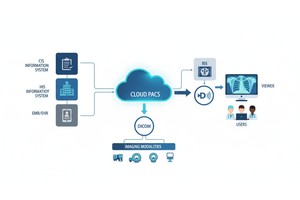
Cardiac computed tomography (CT) or magnetic resonance (MRI) exams are generally stored in DICOM format and then analyzed with PACS software or DICOM viewing software. This is important, as often heart imaging involves radiologists, cardiologists, surgeons, and referring physicians.
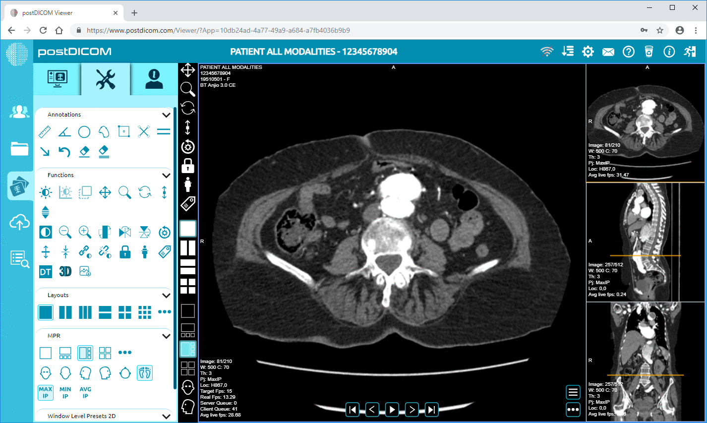
The online DICOM viewer for CT and MRI will help doctors interpret studies, compare findings, and use advanced visualization tools, while also bypassing the need for local workstations.
Access is also essential when a patient needs referral, a second opinion, and/or a multi-disciplinary review. When that occurs, physicians could be required to share cardiac CT and MRI scans securely with other members of the healthcare team in a secure manner.
Cardiac CT is usually preferred if the main concern is the coronary arteries (for example, if the coronary arteries are narrowing, or if there is any doubt about coronary artery disease).
Cardiac MRI is generally best when the primary concern is the heart muscle (how it is working, whether it is inflamed, scarred, damaged, or viable).
In a modern cardiac imaging world, the best scan will be selected depending on the clinical question. When these tests are used appropriately, they assist the physician in the diagnosis, treatment planning, and care decision-making for heart disease.
Cardiac MRI is more suitable for assessing heart muscle function, inflammation, scarring, and damage. Cardiac CT can provide better information about the coronary arteries, plaque, calcium deposits, and constricted blood vessels. The better test depends on the conditions the doctor would like to diagnose.
Cardiac CT is typically preferred for evaluating the coronary arteries and detecting plaque, plaque calcification, or narrowing. Cardiac MRI can help demonstrate the effects of reduced blood flow on the heart muscle, while CT is typically used to directly visualize narrowed or blocked coronary arteries.
A cardiac MRI may demonstrate decreased blood flow, abnormal heart muscle function, scarring, or poor heart function, which may be associated with coronary artery disease. It is not typically the first imaging test used to directly examine coronary artery blockages, however.
Cardiac MRI does not involve ionizing radiation, whereas cardiac CT does. But the patient has to be safe. Certain individuals may not be able to undergo MRI because of implants or devices, or because CT contrast may be contraindicated in patients with kidney disease or a contrast allergy.
Yes. Cardiac CT and cardiac MRI can be used together. CT can be used to assess the coronary arteries, and MRI can be used later to assess damage, inflammation, scarring, or viability of the heart muscle.
|
Cloud PACS and Online DICOM ViewerUpload DICOM images and clinical documents to PostDICOM servers. Store, view, collaborate, and share your medical imaging files. |