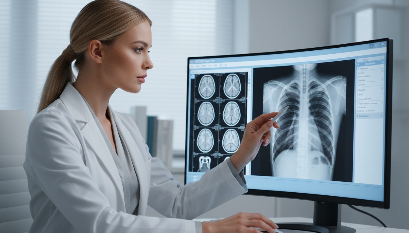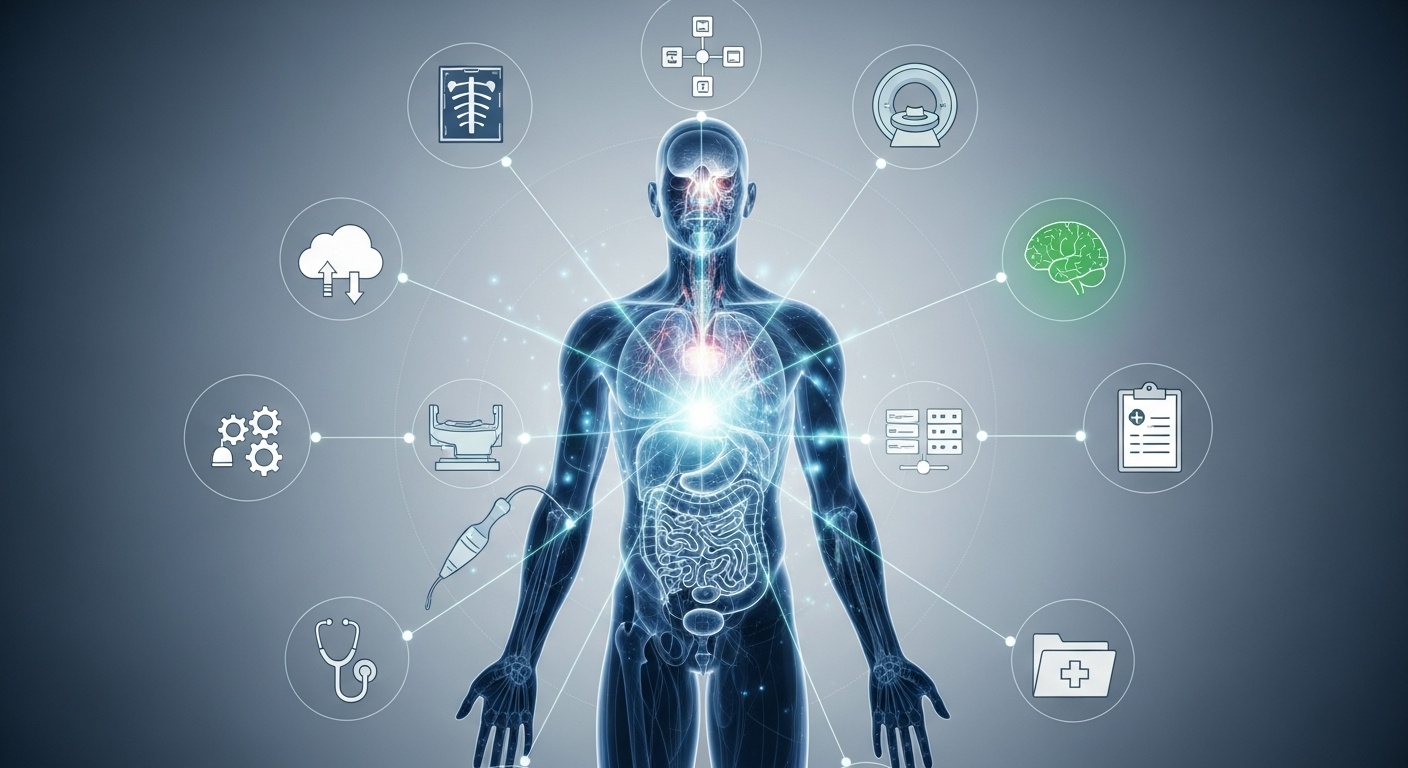
Diagnostic medical imaging has revolutionized the way medical professionals evaluate disease, plan treatment, monitor progress and coordinate care teams. Each new development has improved the clarity, access to, and sharing of medical images—from X-ray to cloud-based imaging workflows. Diagnostic imaging today isn't just a matter of taking pictures of the body. It is also reliant on standards, software, secure storage systems and tools that assist clinicians to work more efficiently.
These advances should be used in addition to clinical judgment. The best imaging technique to use, depends on the clinical question, the patient's condition, the type of technology available, and the physician's recommendation.
Diagnostic medical imaging has revolutionized the medical field by enabling healthcare providers to see inside the patient's body, understand their symptoms, assist in the diagnosis process, plan their treatment, and follow up with them over time.
Some of the significant developments are the introduction of the X-ray, CT, MRI, ultrasound, nuclear medicine, DICOM, PACS, 3D reconstruction, cloud image sharing, and AI-supported imaging workflows. There are different roles for each technology. There are many that are used for soft tissue assessment, some that can be used in real-time imaging, and some that can be used to securely store, view, or share imaging studies.
All of this has helped to create a more useful, more connected and faster imaging workflow.
Diagnostic medical imaging is the use of technologies to produce images of the internal body structures and functions. These images can be used by doctors and other health care providers to look for symptoms, to confirm their diagnosis, to plan a procedure, and to monitor the changes over time.
Typical diagnostic imaging techniques are X-rays, computed tomography (CT), magnetic resonance imaging (MRI), ultrasonography, mammography, fluoroscopy, positron emission tomography (PET) and single-photon emission computed tomography (SPECT). The purpose of each modality is different. Sometimes, X-rays are used for bones or for imaging the chest. CT can offer detailed cross-sectional images. In some situations, MRI may be preferred for certain evaluations of soft tissue. When real-time imaging and no ionizing radiation is required, ultrasound can be used.
The imaging technique is selected by the doctor depending on the evaluation that needs to be performed. For instance, the ideal method for evaluating a fracture might not be suitable for evaluating soft tissue, blood supply, or metabolic activity.
One of the first big innovations in medical imaging was the X-ray image. It enabled doctors to study the bones, lungs and some internal organs in a non-surgical manner. Despite the availability of new technologies, x-rays are still used for many routine checks, and it is quick and easy to get.
X-ray imaging might be conducted in an emergency department, when someone visits the dentist, in orthopedic assessment, chest imaging, or as a follow-up examination. The use of an X-ray, however, will depend upon the patient's symptoms, medical history, and the doctor's evaluation.
Clinicians' views were widened by CT and MRI. CT scanning is a technique that produces cross-sectional images and can be used in trauma evaluation, emergency care, cancer staging, and vascular assessment. MRI makes use of magnetic fields and radio waves to produce detailed images, and is often used to examine the brain, spine, joints, and soft tissues.
Neither of the two scans is superior in every aspect. CT might be the preferred imaging technique if speed is important or if bone or chest detail is required, and MRI may be preferred if detailed soft-tissue information is required. This will depend on the clinical question, its urgency, the patient's clinical status, and the type of information the doctor requires.
Ultrasound works by sending out sound waves to produce an image in real time. It is a commonly used modality for pregnancy, abdominal imaging, vascular imaging, cardiac imaging, and emergency medicine. Ultrasound imaging can be used to observe motion in real time, assist with procedures, and assess organs at the patient's bedside.
The utility of ultrasound often lies in its portability and non-ionizing nature. However, the image's quality and appropriateness will depend on the body area being examined, the patient's condition, and the clinical purpose.
Functional imaging methods (e.g., PET and SPECT) reveal the function of tissues and organs. These do not need to show only structure; they can show information on metabolic activity, blood flow, or organ function. This can be applicable in oncology, cardiology, neurology, and nuclear medicine, depending on the patient's clinical situation.
By combining anatomic and functional data in a single study, hybrid imaging (e.g., PET/CT) may provide a more comprehensive picture to the clinician. These scans are typically chosen when functional data may be useful to answer a particular clinical question.
 - Created by PostDICOM.jpg)
With the advent of digital imaging from film, the healthcare organization required a secure method of storing, transmitting, and displaying images. The DICOM standard in medical imaging helped solve this by standardizing medical image files and related metadata.
DICOM makes it easier for imaging devices, viewing software, and storage systems to communicate. This is important because medical images frequently need to be transferred from one scanner to another, to the radiologist, to the referring physician, to the hospital, and to the clinic.
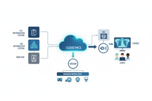
Picture Archiving and Communication System (PACS) revolutionized the way images were stored and retrieved. Healthcare organizations can store and retrieve digital studies, rather than relying on physical films. Today's cloud PACS provide access to the cloud, storage scalability, and support for collaboration among radiologists, physicians, clinics, and hospitals.
| Imaging advancement | Main purpose | Healthcare impact |
| X-ray | Basic internal imaging | Made non-invasive evaluation more accessible |
| CT | Cross-sectional imaging | May support trauma, cancer, and emergency assessment |
| MRI | Detailed soft tissue imaging | Often useful for brain, spine, joint, and soft tissue evaluation |
| Ultrasound | Real-time imaging | May support bedside imaging and guided procedures |
| PET/SPECT | Functional imaging | May add metabolic and organ-function information |
| DICOM | Image standardization | Improved compatibility between imaging systems |
| PACS | Digital image storage | Replaced film archives and improved image access |
| 3D reconstruction | Advanced visualization | May help with surgical planning and complex anatomy review |
| Cloud imaging | Remote access and collaboration | May support image sharing across locations |
| AI-supported workflows | Workflow and image analysis support | May assist with prioritization, segmentation, or quality control |
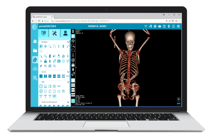
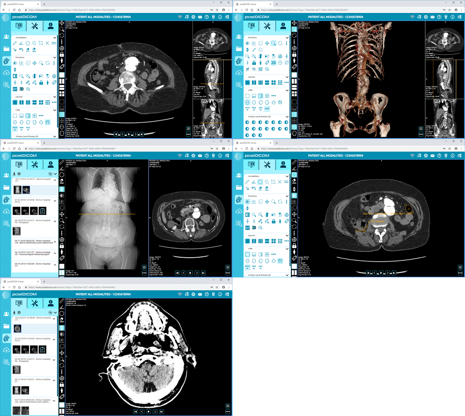
The imaging process doesn't end once the scan is taken. A post-processing tool assists the clinician in better displaying and interpreting images. Sometimes, complex anatomy may be easier to understand with techniques such as multiplanar reconstruction, maximum-intensity projection, minimum-intensity projection, and 3D reconstruction.
These workflows can be handled by a Diagnostic DICOM Viewer, which can assist users with the following functionalities:
Clinicians use advanced viewing tools extensively whenever they must examine anatomy at various angles, compare studies or make images for consultation. The usefulness of these tools will be dependent on the clinical setting and the type of imaging study being considered.
Many areas of healthcare use diagnostic imaging. In emergency medicine, CT, X-ray, and ultrasound may be used to aid the diagnosis of trauma, stroke symptoms, internal bleeding, and acute pain. The mode of delivery depends on the urgency of the situation and the doctor's requirements.
CT, MRI, PET/CT, and nuclear medicine imaging can be useful in oncology for the detection, staging, treatment planning, and follow-up of tumors. These imaging techniques can also aid in tracking changes over time, but only within the context of the patient's clinical situation.
In the field of cardiology, an echo, computed tomography (CT) angiography, magnetic resonance (MRI), and nuclear imaging might be useful to assess the structure, flow, and function of the heart. Many orthopedic teams rely on these technologies to evaluate fractures, joints, ligaments, and spine conditions. In a remote-care scenario, digital imaging workflows can simplify radiologists' and other specialists' ability to review studies from remote locations.
A major shift in diagnostic imaging is from isolated systems to connected digital workflows. The typical workflow of an imaging system could involve image acquisition, DICOM conversion, PACS storage, diagnostic viewing, reporting, sharing, and long-term archive.
Secure medical image sharing is especially useful when patients need referrals, second opinions, specialist review, or follow-up care at another facility. Digital sharing can provide quick, easy access for authorized users without the need for a CD or physical film.
Cloud-based imaging platforms can also assist smaller clinics, imaging centers, and distributed healthcare teams to handle imaging information without the need of huge on-site infrastructure. This is beneficial for businesses that require remote access, collaboration, and scalability.
Medical imaging workflows are increasingly becoming visible with the help of AI. AI tools can be used for image prioritization and to assist with image detection, segmentation, quality control, and workflow automation.
AI is not a substitute for medical expertise, though. Imaging results must be interpreted in the context of the clinical situation, and a thorough review and analysis of these images must be performed by a trained health care professional. AI's biggest contribution is as a supporting tool in a secure, vetted, and controlled imaging process.
AI tools also must be carefully assessed by healthcare organizations. There are several key considerations for AI in healthcare settings, including accuracy, workflow integration, data quality, regulatory compliance, and human oversight.
Although significant strides have been made, some challenges remain in diagnostic imaging. High-tech scanners and image software can be costly. If you use a lot of large photos, you need a safe place to store them and access them quickly. Robust privacy and security measures must also be in place for healthcare organizations to safeguard patient information.
Interoperability issues with systems, staff training, data migration issues, and workflow consistency across departments/locations are other challenges. In an increasingly digital and connected world of imaging, healthcare professionals must be equipped with healthcare information systems that benefit both clinical effectiveness and responsible data management.
Key developments include X-ray, computed tomography (CT), magnetic resonance imaging (MRI), ultrasound, PET/CT, DICOM, PACS, 3D reconstruction, cloud imaging, and AI-driven imaging workflows. Some improvements enhanced image capture, while others enhanced image storage, viewing, sharing, and analysis. They have made imaging more useful for diagnosis, treatment planning, follow-up, and collaboration.
Diagnostic imaging might be used to look inside the body, to identify abnormal areas, to guide procedures, to plan treatment, and to track changes over time. It can help assess some conditions at an earlier stage than they would otherwise be evaluated, and it can minimize the need for certain exploratory procedures. Imaging is not the whole story, though. Diagnosing images along with symptoms, physical exam, laboratory results, and medical history.
The significance of DICOM is that it provides a uniform representation of medical images and information among imaging devices and imaging software. Imagine if there were no standard, such as DICOM, and hospitals had to carry out imaging studies in a new way. If DICOM did not exist, how would it be easier for scanners, PACS software, scanner viewers, and hospitals to reliably exchange imaging studies? DICOM also supports significant metadata, such as study information, modality type, image information, etc.
Medical image files and communication are standardized by DICOM. Those images are stored, retrieved, managed, and distributed by a system known as PACS. The difference can be likened to the way photos are captured and conveyed versus how they are stored and handled by healthcare staff. The distinction is easy to make: DICOM describes how image data is structured and transmitted, and PACS is the archive and workflow system that enables healthcare staff to access and work with images.
AI is mainly employed as an assisting tool in the imaging workflow. It can aid in prioritizing studies, pattern recognition, segmenting anatomy, or enhancing workflow efficiency. The role of the radiologist and any qualified health care professional continues to be the interpretation of the image within the correct clinical context. AI-generated content should be thoroughly evaluated and should not be used as a substitute for a diagnosis.
Cloud imaging can be used to enable remote access, image sharing, collaboration, and scalable storage. It can be helpful to hospitals, imaging centers, clinics and teleradiology teams that require authorized users to view studies from different locations. Benefits depend on proper implementation, secure access controls, privacy protection, and integration with the organization's current imaging workflow.
|
Cloud PACS and Online DICOM ViewerUpload DICOM images and clinical documents to PostDICOM servers. Store, view, collaborate, and share your medical imaging files. |